(Submitted by CHS Junior, Cali Hendrickson)
Sheep Brain Dissection:
Mr. Bradley’s Anatomy class worked on an exciting journey into the inner workings of the brain with a hands-on sheep brain dissection. The objective was to identify and describe the principal structures of the sheep brain. This engaging activity offered students an opportunity to observe the complexity of the nervous system firsthand.
Examination of the Sheep Brain
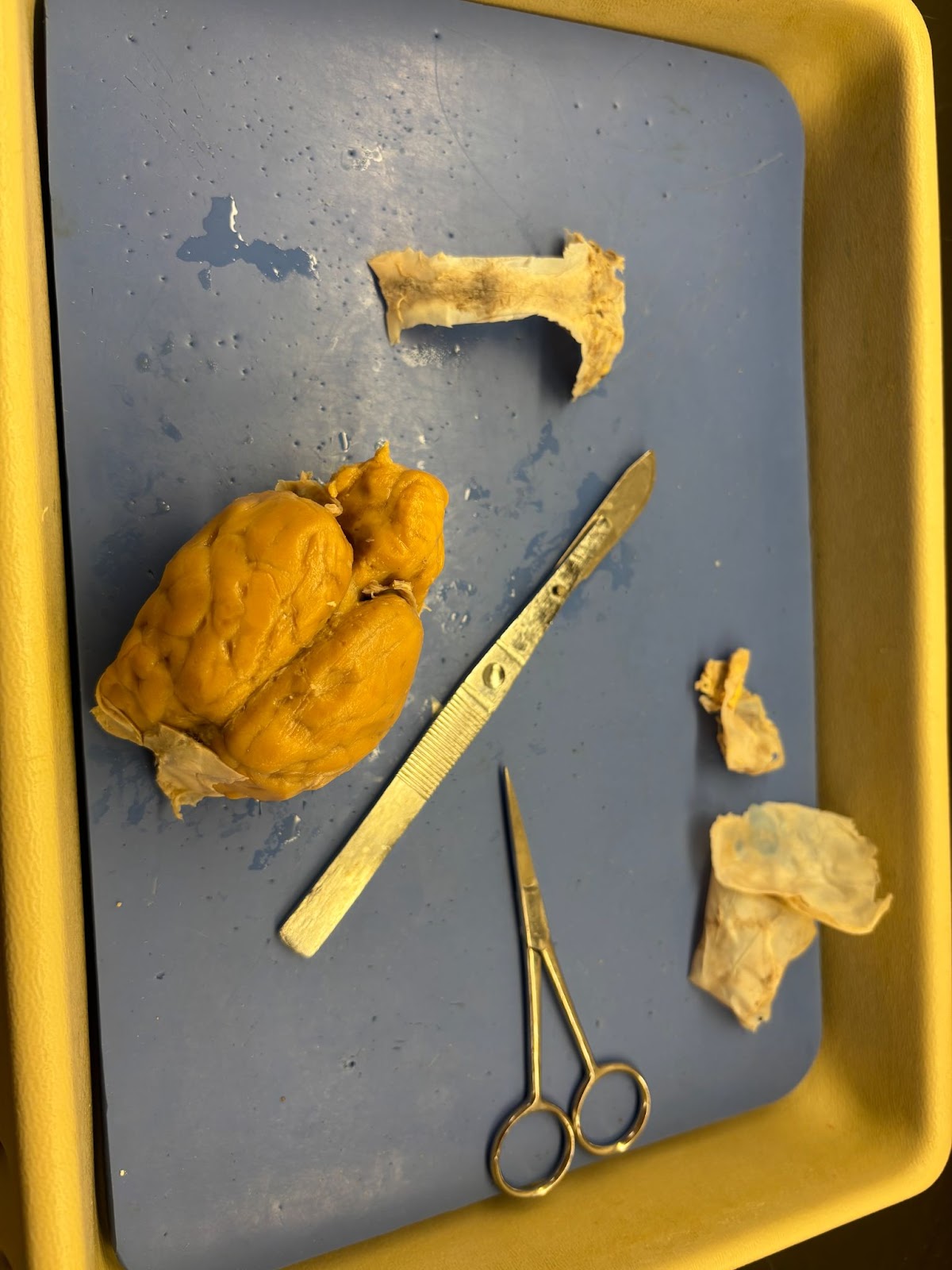
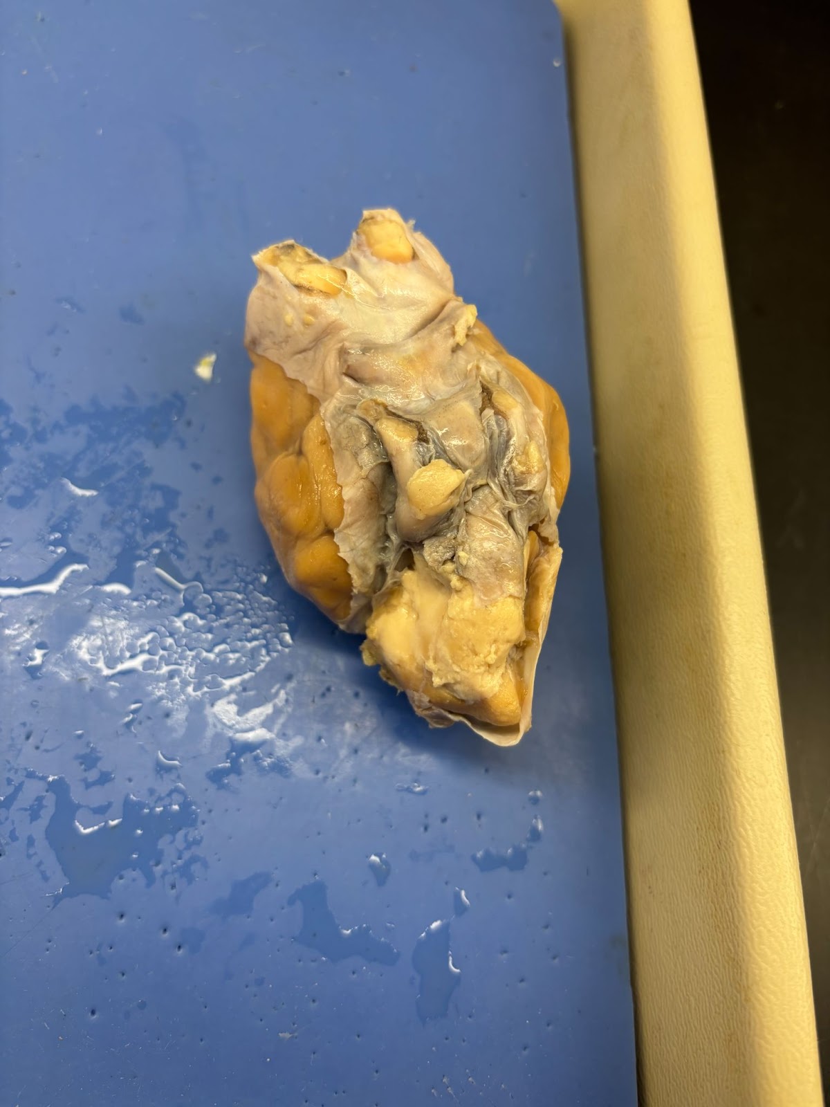
The dissection began with an external examination of the sheep brain. Interestingly, the sheep brain is quite similar to the human brain in structure but differs in size and orientation. The sheep brain is elongated with an anterior-to-posterior orientation, while the human brain is positioned more vertically.
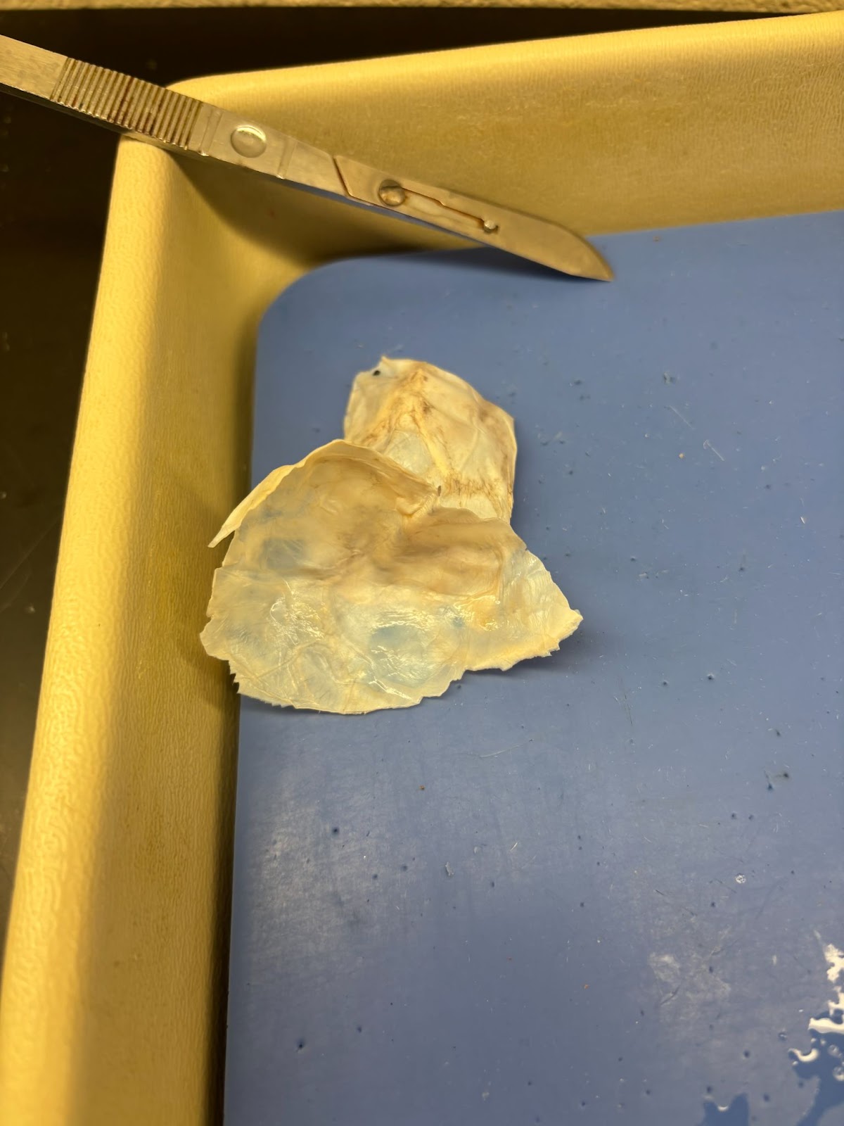
Dura Mater Removal
The tough outer covering of the brain, known as the dura mater, was carefully removed. This membrane protects the brain but must be taken off to view the underlying structures.
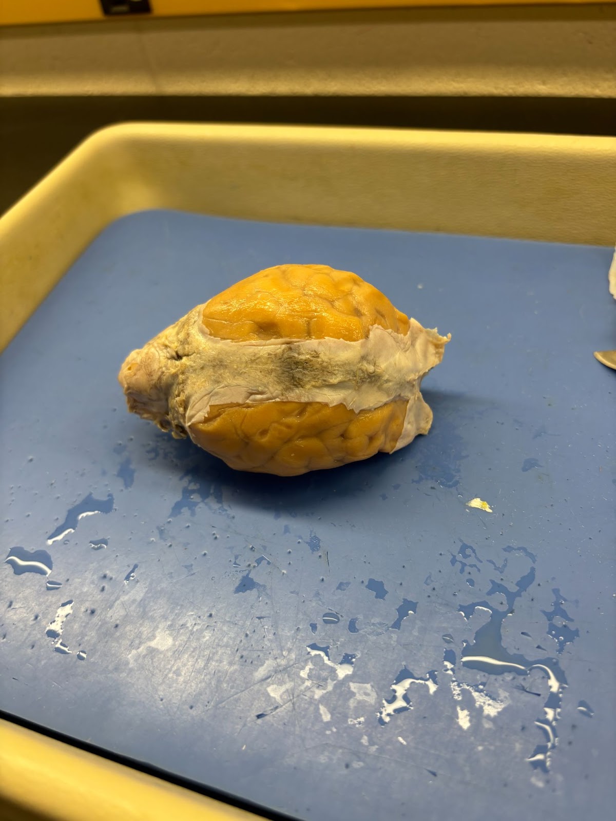
Identification of the Cerebrum
The cerebrum, the most prominent part of the brain, was observed. It is divided into two nearly symmetrical hemispheres by the deep longitudinal fissure.
Observation of the Gyrus and Sulcus
The surface of the cerebrum is covered in folds known as gyri and grooves called sulci. These folds increase the surface area of the brain, allowing for more complex neural connections.
Cerebellum Examination
At the back of the brain lies the cerebellum, responsible for balance and coordination. Its smaller gyri distinguish it from the cerebrum.
Exploring the Ventral Surface
Turning the brain over revealed several crucial structures:
-Optic Chiasma: An X-shaped structure where optic nerves cross, crucial for vision.
-Pituitary Gland: Although often removed with the dura mater, its location below the optic chiasma was noted.
-Olfactory Bulbs: Located at the front of the brain, responsible for the sense of smell.
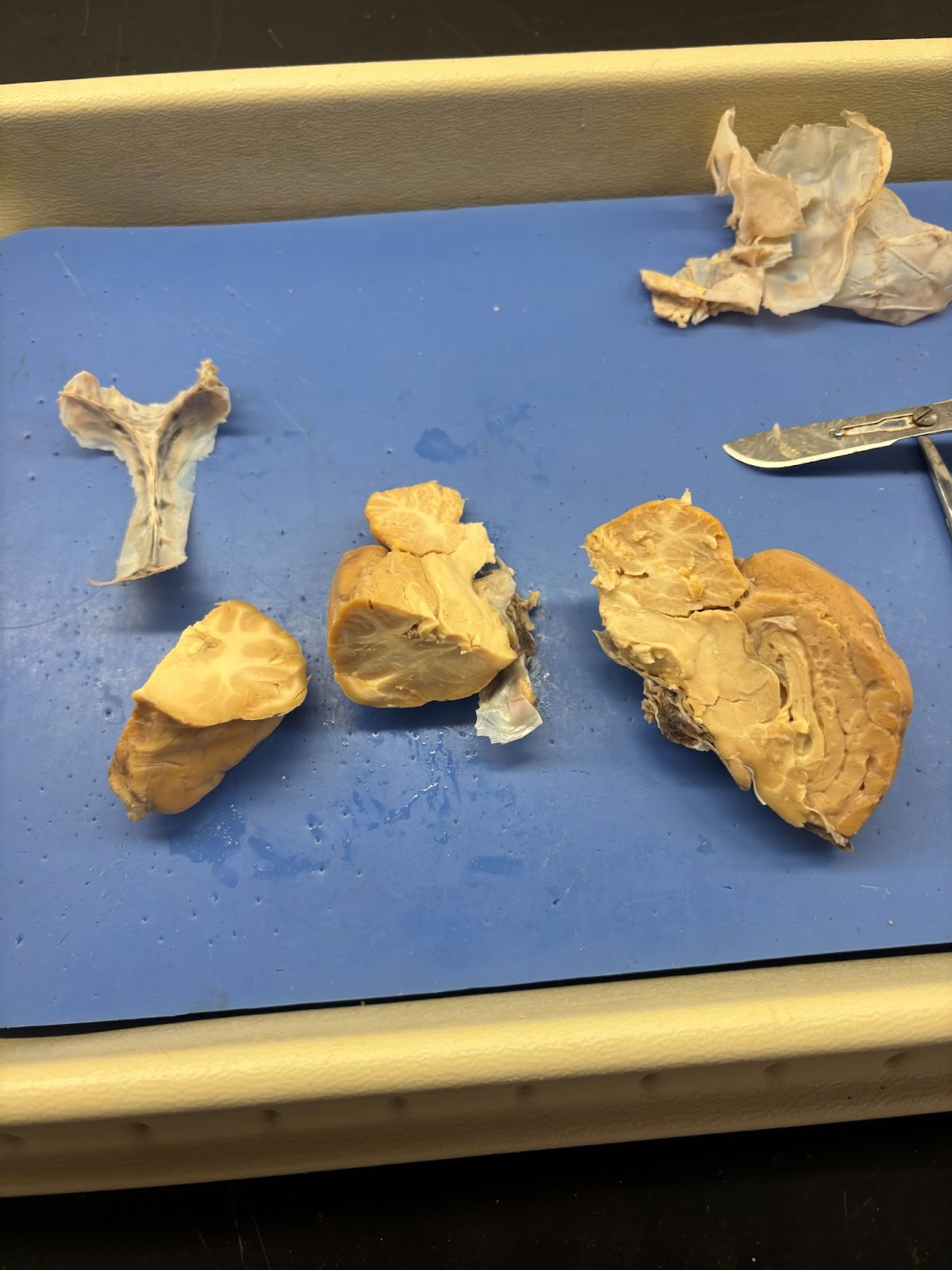
Dissection of the Internal Brain
A longitudinal incision along the deep longitudinal fissure was made to examine the internal structures to separate the brain into the left and right hemispheres. This revealed:
Corpus Callosum
The bundle of nerve fibers connecting the two cerebral hemispheres is crucial for communication between the brain’s halves.
Thalamus and Hypothalamus
These structures were visible beneath the corpus callosum. The thalamus acts as a relay center for sensory signals, while the hypothalamus regulates vital bodily functions like temperature and hunger.
Pineal Gland and Third Ventricle
The pineal gland, involved in regulating sleep cycles, was observed near the third ventricle, a cavity containing cerebrospinal fluid.
Conclusion
Mr. Bradley’s Anatomy class successfully completed the sheep brain dissection, gaining a deeper understanding of the brain’s intricate design and functionality. This hands-on experience not only enhanced their knowledge but also sparked curiosity about the wonders of neuroscience.
Stay tuned for more exciting explorations in Anatomy from Mr. Bradley’s classroom!