 (Submitted by CHS Senior, Alea Brierly)
In Mr. Bradley’s third block anatomy and physiology class, we have been learning about the anatomy and physiology of the cardiovascular system. Along with the blood typing lab, we dissected a pig heart. We have been reading and listening to lectures about how the heart works, the different valves and vessels, the circulation but there’s no better way to learn then to dissect something and see it for yourself.
After we rinsed the heart all off, we had to remove the pericardium, the outer sac that surrounds the heart. We located the apex, which is the tip of the heart. Before cutting the heart in half, we located several chambers and blood vessels including, the left and right atria, left and right ventricles, the coronary artery which supplies the heart with blood, pulmonary artery which brings deoxygenated blood to the lungs to get oxygen, aorta which sends blood out to the body and pulmonary veins which bring oxygenated blood back into the left atrium.
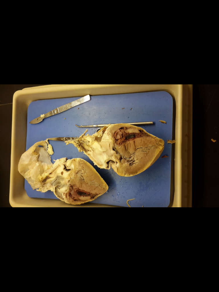
 We then cut the heart in half to look at the internal anatomy. We located the same things that we did on the outside but we also looked at the tricuspid valve which is located between the right atrium and the right ventricle. The tricuspid valve has three leaflets to prevent the blood from flowing backwards. This valve allows blood flow from the right atrium and the right ventricle during diastole, period where heart is relaxing. The bicuspid valve is between the left atrium and left ventricle. This valve only has two leaflets because it is more important that the blood doesn’t flow back through the right side. This valve is the systolic phase or contracting phase. We found the septum on the right side of the right ventricle. This thick muscular wall separates right and left pumping ventricles. We also found oneway, semilunar valves which only allow blood one direction, these are located at the entrance of the pulmonary veins.
Here’s some pictures of the heart cut open with the pericardium above and the one on the right is Mr. Bradley helping students locate some of the chambers and valves.